

3D visualization of perivascular spaces (PVS) segmented from brain MRI, highlighting their distribution throughout the cerebral white matter.
Neuroimaging
At the Brain Clearance Research Center, we utilize advanced MRI techniques to visualize and quantify brain clearance pathways, offering critical insights into their roles in health and disease. Our team has developed and optimized state-of-the-art imaging methodologies to examine the structural and functional aspects of brain clearance, focusing on perivascular spaces, fluid dynamics, and their relevance to neurological disorders.
The BCRC has been at the forefront of this emerging field, pioneering novel imaging methods and shaping the scientific understanding of brain clearance. Our comprehensive review paper highlights the latest advancements and challenges in brain clearance imaging, reinforcing our leadership in developing new neuroimaging techniques for this critical area of neuroscience.
Below are key imaging tools pioneered by the BCRC:
Structural Imaging of Brain Clearance Pathways (EPC Method)
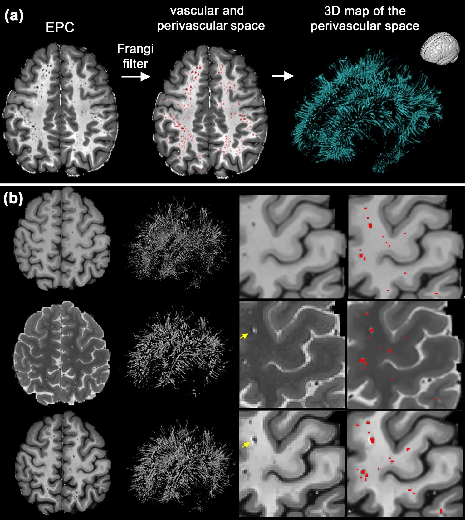
Our team developed the EPC (Enhanced Perivascular Contrast) method, a novel imaging approach that enhances visualization of brain clearance structures by integrating T1- and T2-weighted MRI scans. This technique allows for improved contrast and delineation of perivascular spaces, offering a more detailed assessment of their morphology and potential dysfunction in neurological conditions.
Functional Imaging of Brain Clearance Pathways (TTI Method)
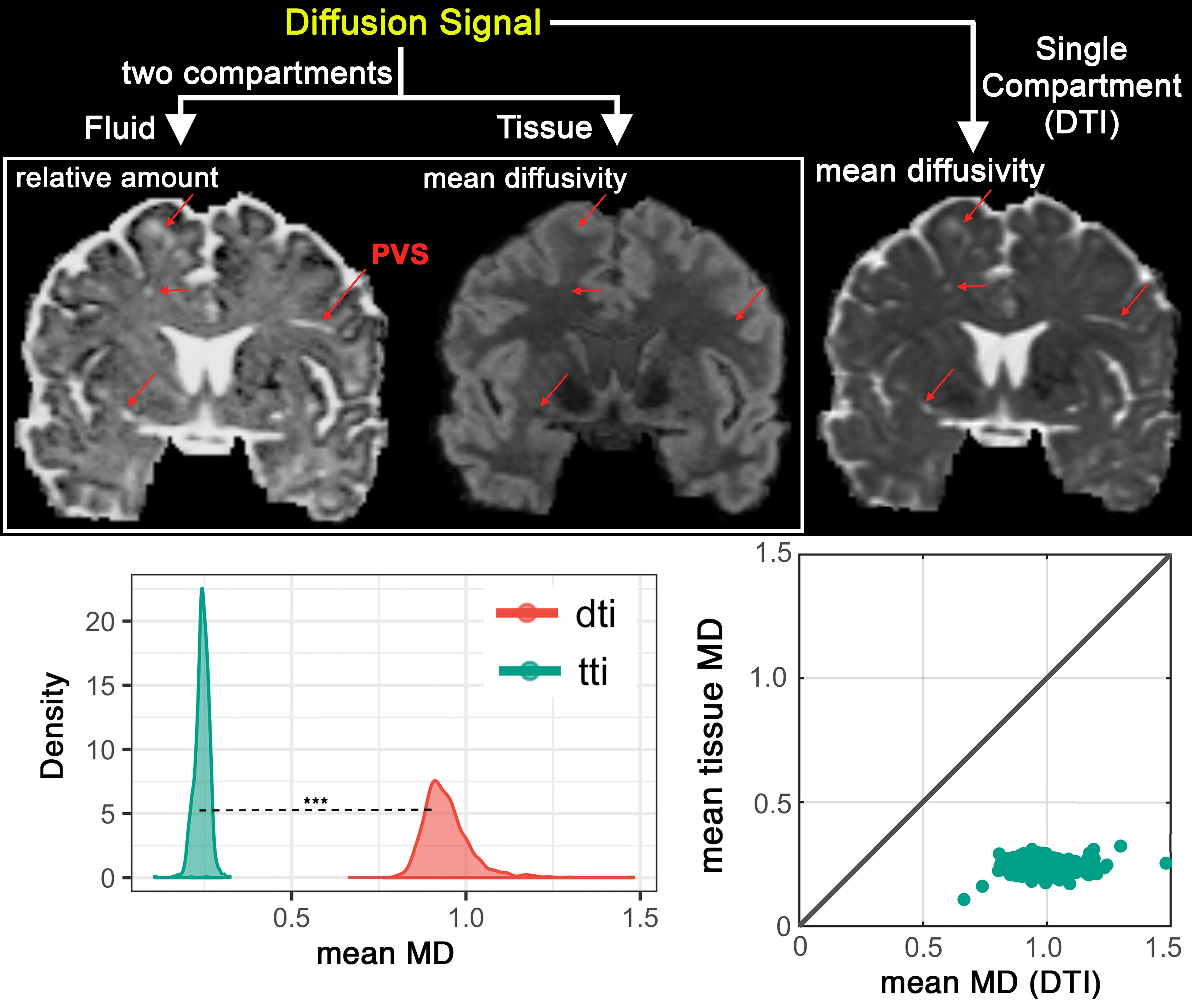
To explore the dynamics of brain clearance, we use diffusion MRI techniques to assess fluid properties within perivascular spaces. Our TTI (Tissue Tensor Imaging) method, derived from bi-tensor modeling of multi-shell diffusion MRI data, enables us to track parenchymal and perivascular space fluid diffusion characteristics, providing a functional perspective on how waste is cleared from the brain. This approach is particularly valuable for understanding clearance impairments in neurodegenerative diseases.
Ultra-High-Resolution Imaging of Perivascular Spaces (7T MRI)
We have pioneered ultra-high field MRI imaging, using the Stevens INI’s 7T MRI scanner, to achieve unprecedented detail in perivascular space imaging. Our advancements allow for visualization of up to 10 times more perivascular spaces than previously possible, shedding light on their microstructural organization and potential alterations in conditions like Alzheimer’s disease.
Multi-Modal Clinical Imaging for Neurological Disorders
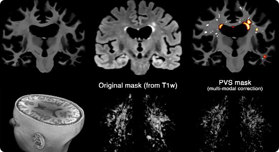
In our efforts to translate neuroimaging advancements to clinical applications, we have developed a multi-modal imaging protocol combining T1-weighted, T2-weighted, and FLAIR MRI sequences. This integrative approach enhances our ability to assess brain clearance dysfunction in neurological disorders, particularly Alzheimer’s disease, where impaired waste clearance may play a crucial role in disease progression.
Deep Learning for Automated Mapping of Perivascular Spaces
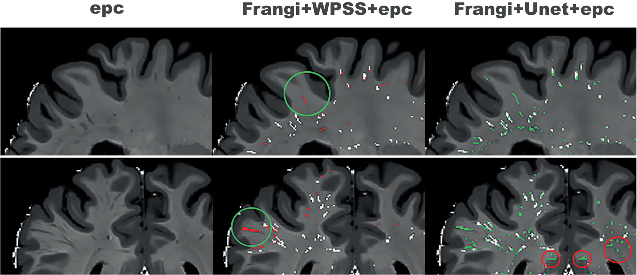
Leveraging artificial intelligence, we have developed a deep learning-based method for automated mapping of perivascular spaces (PVS) from MRI scans. This cutting-edge approach enables highly accurate, large-scale quantification of PVS, reducing manual segmentation efforts while improving consistency and precision. By harnessing deep neural networks, our method enhances the study of brain clearance dysfunctions and facilitates biomarker discovery for neurological diseases.
Through these innovations, the Brain Clearance Research Center is at the forefront of neuroimaging research, providing valuable insights into brain clearance mechanisms and their role in neurological health.